


To understand why floaters and flashes happen, you first need to review a little bit of eye anatomy.
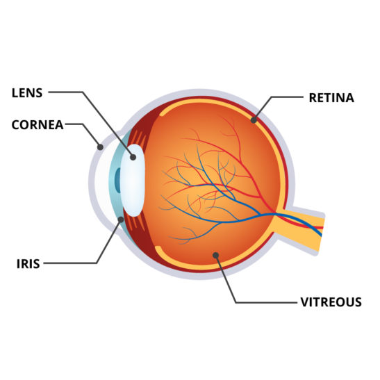
The eye is essentially a ball which I like to divide into three areas of interest. The front (or cornea) is the window that allows light into the eye. The middle is composed of the iris, pupil and focusing lens. Behind the iris-lens area is a large cavity filled with a clear gel-like substance called vitreous. And behind the gel is the back wall that is lined with a thin “wallpaper” called the retina.
Most do not realize that the retina is actually an extension of the brain. So retina is really brain tissue! It is responsible for transforming light that enters the eye into electrical signals that are processed and interpreted by the rest of the brain into shape and color. You really do not see with your eye. The eye is really a transducer of energy, light into electrical signals. The rest of your brain forms the image.
- Anatomy of an Eye
Why is the doctor so interested in FLASHES and FLOATERS?
The usual normal process of vitreous gel aging and separation from the retina occurs over a lifetime and does not usually cause problems. The gel liquefies and collapses separating from the retina.
During this process, the patient may begin to notice small translucent or darker particles occasionally “floating” in their field of view. These floaters represent the projected shadows of fine vitreous particles floating in front of the retina. In some situations, however, the separation of the gel occurs too suddenly or the pull of the gel is too forceful for the delicate retina resulting in retinal tears.
Usually, these more violent vitreous separations are associated with more symptoms- more noticeable flashes and floaters. (The retina does not have pain fibers only light fibers so when pulled or torn, light flashes are seen). Doctors take all flashes seriously. With careful examination, it is possible to determine if tears occur and treat damaged areas before any further changes follow.
Often a tear may be associated with many floaters, not just a few minor shadows, but large clumps. These larger and more numerous floaters often represent the clotted blood that exudes from vessels at the edge of the torn retina. These clots absorb over time, the floaters appear to decrease. The tear or tears do not repair themselves and some permit liquid to invade beneath the retina producing first a blister of retinal tissue around the tear(s).
With time and eye movement, more and more fluid infiltrates under the retina creating a larger separation of the retinal “wallpaper”. When larger separations are present they are called retinal detachments. A detached retina is seen by the patient as an increasing shadow or veil-like black area within the normal field of view. Detachments require more extensive treatment (often surgery) to reattach the separated areas.
So floaters may be simply a normal aging process (common) or a symptom of something more ominous. Most individuals go through the process of vitreous gel separation without tear formation, however, it is always prudent to have a careful exam until the gel separation is complete and the symptoms subside.
Strong flashes and many floaters are more alarming symptoms and are more often associated with damage that may require immediate attention. Early detection is the best way to improve the chances for success in treating retinal tears and avoiding detachments with potential serious vision loss.
An examination is always better sooner than later.


