Laser photocoagulation is a treatment most often used for diabetic retinopathy that uses a laser to seal leaking blood vessels. During later stages of diabetic retinopathy, abnormal blood vessels grow in response to damage within the retina. These new capillaries are weak and will often break, leak, or burst. When this happens, sudden and severe vision loss can occur.
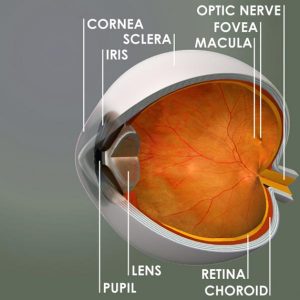
During laser photocoagulation, a laser is used to direct intense, focused rays of light through the transparent structures of the eye to the retina. The laser will burn, or coagulate, the tissues around the abnormal blood vessels, destroying the blood vessels and scarring the tissues so that new blood vessel growth cannot occur. The laser is not directed toward the macula, the area responsible for clear, distinct, straight-ahead vision, as it would destroy sight. Vision is preserved using this procedure but is not restored.
Treatment is usually a short, painless, and relatively easy process, done in the doctor’s office, and does not require hospitalization.
Laser photocoagulation is sometimes used to treat type 2 neovascularization and central serous chorioretinopathy, as well as for detached retinas.
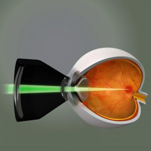
The image below shows how laser light is focused on to specific areas of the retina using a "contact lens" held by the doctor.