Fluorescein angiography (FA) is a technology that allows photographs of the blood vessels of the retina to be taken. It is a useful tool for the diagnosis and treatment of several forms of macular degeneration and retinopathy as well as other disorders.
The process involves injecting the dye into a vein in the arm, just after the eyes have been dilated with eye drops. The yellow dye will travel throughout the bloodstream, including the vessels of the retina. It will fluoresce, or emit light, when an extremely bright light is flashed into the eyes. A series of photographs will show the entrance and exit of the dye through the blood vessels of the retina and just beneath it. Any areas of abnormal new blood vessel growth, leakage, or blockage, will appear darker or brighter than normal angiograms of the vascular retinal area.
The process is generally safe, with a minimum of discomfort, and is usually done in the doctor’s office. It is an important tool, because it will reveal the location of damage by leaking or pooled blood and will show where new blood vessel growth is occurring. This will help the ophthalmologist to decide upon the best treatment to prevent further vision loss.
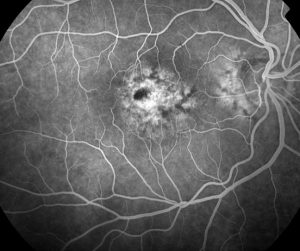
Below is a typical flourescein angiogram showing an eye with leaking vessels (the bright area) in the middle of the macula. The optic nerve is seen to the right.