Indocyanine Green (ICG) Imaging is similar to fluorescein angiography (FA). Like FA, it is a useful tool for the diagnosis and treatment of several forms of macular degeneration and retinopathy. It is more effective than FA in producing images of blood vessel growth and damage in the deeper choroid layer behind the retina. It is also able to produce a better image than FA if blood is present in the macula.
The process is generally safe, with a minimum of discomfort, and is usually done in the doctor’s office. It involves dilating the eyes being photographed, and an injection of the green dye, usually in the arm, which circulates throughout the body, including the choroidal layer of the eye. A flash illuminates the eye and the resulting images are stored on a digital camera system. The fluorescent light that the indocyanine green dye emits can help to identify abnormalities in the circulation of the choroid. A series of images, or a movie of the dye moving through the vessels of the choroid, will help the ophthalmologist to decide upon the best treatment to prevent further vision loss.
Most ophthalmologists consider ICG angiography to be an adjunctive or secondary test which adds further information to the clinical picture, and is often done in addition to a fluorescein angiogram.
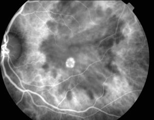
Below is an example of a typical Indocyanine Green angiogram, revealing an area of leakage (the bright area) just outside the macula in the center of the image. The optic nerve can be seen to the left.